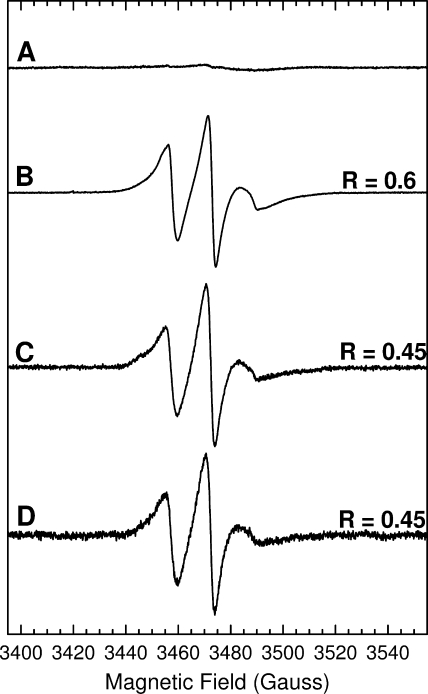
FIGURE 1.
Comparison of the effects of different spin dilution methods on the room temperature X-band EPR spectra of the spin-labeled TatA Ile15 → Cys variant. A, wild-type TatA at a protein concentration of 1 mm. B, 100% labeled Ile15 → Cys TatA. The protein concentration was 350 μm. C, 10% labeled Ile15 → Cys TatA prepared by sub-stoichiometric addition of MTSL. The protein concentration was 800 μm. D, 10% labeled Ile15 → Cys TatA prepared by dilution of 100% labeled Ile15 → Cys with wild-type TatA. The final protein concentration was 350 μm. Spectral intensities have been normalized for comparison of line shapes. Spectra were recorded under the following conditions: frequency = 9.7 GHz, power = 2.0 milliwatts, modulation amplitude = 1 gauss, temperature = 295 K. R is the ratio of the low field peak height to that of the central peak.

