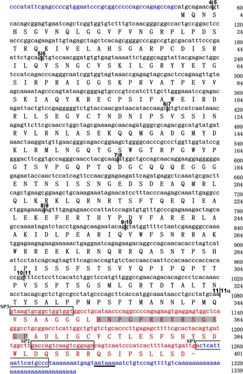
FIGURE 2.
The nucleotide and protein sequences of full-length Pax6(S). Nucleotides are in lowercase letters, and amino acids are capitalized. Boundaries between exons are labeled by vertical lines with numbers on both sides indicating the preceding and following exons, separately. Exons are numbered the same as in Pax6 gene (except for exon 11α). Exon 11α and the S tail are in red. The cDNA sequences to which the SP1, SP2, and SP3 primers annealed (see “Experimental Procedures”) are framed. The epitope that the Pax6(S) antibody recognizes is in shadow. 5′- and 3′-UTRs are in blue. The polyadenylation signal in the 3′-UTR is underlined.

