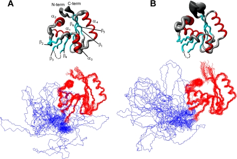
FIGURE 1.
Solution structures of ATP-bound N-MNK (A) and ATP-free N-MNK (B). The average backbone RMSD between ATP-free and ATP-bound N-MNK domains is of 1.01 Å. The secondary structure elements comprise residues 1054–1061 (β1), 1070–1081 (α1), 1090–1100 (α2), 1108–1114 (β2), 1118–1124 (β3), 1178–1183 (β4), 1186–1190 (α3), 1197–1209 (α4), 1212–1218 (β5), and 1221–1230 (β6) for ATP-bound N-MNK and residues 1054–1061 (β1), 1070–1080 (α1), 1087–1100 (α2), 1110–1114 (β2), 1118–1123 (β3), 1178–1183 (β4), 1186–1191 (α3), 1197–1209 (α4), 1212–1218 (β5), and 1221–1230 (β6) for ATP-free N-MNK, respectively. Top panels, the radius of the tubes is proportional to the backbone RMSD of each residue. The unstructured loop was omitted for simplicity. Bottom panel, the backbone traces for the twenty lowest energy conformers are superimposed. The unstructured loop is shown in blue.

