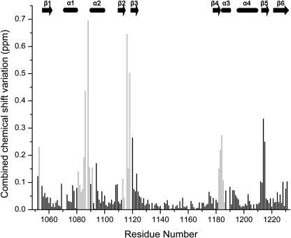
FIGURE 3.
Combined chemical shift variations of signals between ATP-free N-MNK and ATP-free N-MNK in the presence of 3.0 equivalents of ATP. Empty columns correspond to residues whose backbone experienced significant structural changes upon ATP binding; these residues are shown in red/pink in Fig. 5B. Combined chemical shift variations are calculated from the experimental 1H and 15N chemical shift changes (Δδ(1H) and Δδ(15N), respectively) between corresponding peaks in the two forms, through the following equation (43), . The missing assignment for residue Ile1119 of the ATP-free N-MNK prevented the determination of its chemical shifts perturbations. The position of secondary structure elements is shown.

