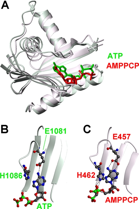
FIGURE 8.
Comparison of the ATP-bound structure of N-MNK and of the N-domain of AMPPCP-bound A. fulgidus CopA showing in detail the protein-substrate interactions. A, superimposition of the overall structures of the N-domain of CopA (molecule A in 3A1C, red) and N-MNK (green). The substrates are shown as sticks. The unstructured loop of N-MNK was omitted for simplicity. Details of the nucleotide-binding regions are shown in B for N-MNK and in C for CopA.

