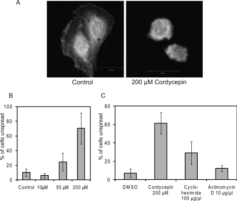
FIGURE 3.
Cordycepin inhibits cell spreading. NIH3T3 cells were detached, suspended for 1 h in serum-free medium, and allowed to re-attach to coverslips with serum for 5 h in the presence or absence of cordycepin, cycloheximide, or actinomycin D at the concentrations indicated. After fixation, cells were stained with phalloidin to visualize the actin cytoskeleton. A, images of typical control and cordycepin-treated cells. B and C, quantitation of the percentage of unspread cells (largest diameter, 25 μm or less) in cells incubated with the indicated doses of drugs.

