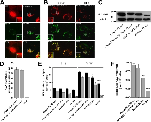
FIGURE 6.
The N terminus of FAAH-2 is a lipid droplet localization sequence. A, COS-7 cells transfected with RFP or FAAH2(N)-RFP were fixed and stained with BODIPY493/503. The top panel shows RFP, the middle panel depicts BODIPY493/503, and the bottom panel shows the merged images. Note that RFP aggregates were present in every FAAH2(N)-RFP-expressing cell examined. B, co-localization of FAAH2(N)-rΔTMFAAH-FLAG with BODIPY493/503 in COS-7 and HeLa cells. C, expression of rFAAH-FLAG, rFAAH-FLAG(low), FAAH2(N)-rΔTMFAAH-FLAG, and FAAH2-FLAG in HeLa cells. rFAAH-FLAG(low) represents rat FAAH-FLAG expressed at levels comparable to FAAH2(N)-rΔTMFAAH-FLAG. The proteins were resolved by SDS-PAGE and probed with anti-FLAG or anti-β-actin antibodies. D, similar (p > 0.05) enzymatic activities of rFAAH-FLAG(low) and FAAH2(N)-rΔTMFAAH-FLAG in homogenates of HeLa cells (n = 3). E, uptake (black bar) and hydrolysis (gray bar) of 100 nm [14C]AEA by HeLa cells transiently transfected with rFAAH-FLAG(low), FAAH2(N)-rΔTMFAAH-FLAG, FAAH2-FLAG, or vector controls following 1- or 5-min incubations. Statistical significance was determined between transfected cells and rFAAH-FLAG(low)-transfected controls. *, p < 0.05; **, p < 0.01; ***, p < 0.001 (n = 3). F, intracellular hydrolysis of [14C]AEA in transfected HeLa cells following uptake at 3 s. **, p < 0.01; ***, p < 0.001 (n = 3).

