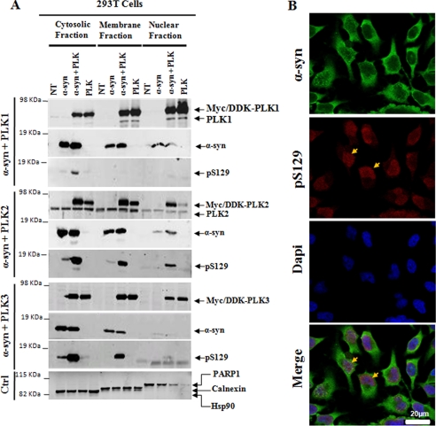
FIGURE 7.
Subcellular localization of α-syn and PLKs in mammalian cell. A, HEK 293T cells were co-transfected with pAAV-CMV-α-syn-WT and different pCMV6-Èntry-PLKs and then subjected to subcellular fractionation 24–48 h post-transfection with the ProteoExtract® subcellular proteome extraction kit from Calbiochem. Purified fractions were then immunoblotted with corresponding antibodies. PARP1, Hsp90, and calnexin proteins were used as control for the nuclear, cytosolic, and membrane particulate fractions, respectively. B, confocal microscopy of HeLa cells co-stained with anti-Ser(P)-129 and anti-α-syn antibodies. Endogenous Ser(P)-129 (red) accumulate exclusively in the nucleus stained with 4′-6′-diamidino-2-phenylindole (Dapi), whereas α-syn WT (green) is mostly membrane and cytosolic in non-transfected cells (NT).

