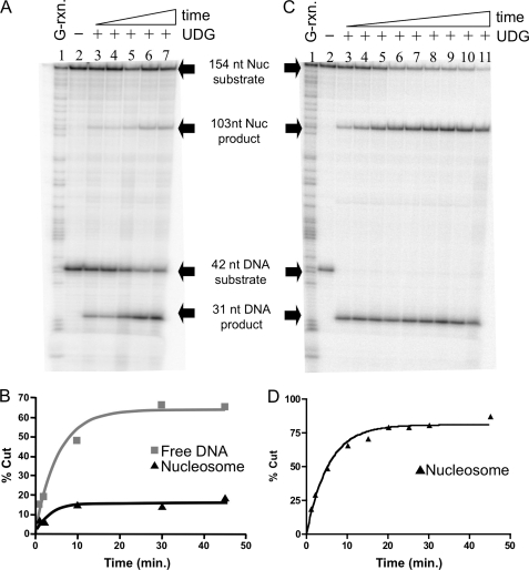
FIGURE 3.
Uracil at an inward facing site within the nucleosome is excised very slowly by UDG. Nucleosomes reconstituted with the 154(+28U) DNA template were combined with the 42-bp reference DNA fragment before incubation with UDG and analysis of cleavage as described in the text. A, nucleosomes and naked DNA were incubated with 0.05 units of UDG. Lane 1, G-reaction marker; lane 2, substrates incubated in the absence of UDG; lanes 3–7, substrates incubated in the presence of UDG for 1, 2, 10, 20, and 30 min, respectively. Substrates and products are indicated as described in the legend to Fig. 2A. B, quantification of data shown in A. C, nucleosomes and naked DNA cleaved with 23.4 units of UDG. Lane 1, G-reaction marker; lane 2, substrates incubated in the absence of UDG; lanes 3–11, substrates incubated for 1, 2, 5, 10, 15, 20, 25, 30, and 45 min, respectively. D, quantification of data shown in C. Data were fitted to a single exponential as described in the legend to Fig. 2B.

