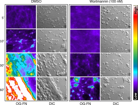
FIGURE 5.
Microscopic analysis of the role of PI3K in platelet-mediated clot retraction. Washed platelets were pretreated with either vehicle (DMSO) or wortmannin (100 nm) and supplemented with 0.25 mg/ml fibrinogen containing 10% Oregon Green-labeled fibrinogen (OG-FGN). Platelets were stimulated with 1 unit/ml thrombin, and the process of clot retraction was visualized in real time using both DIC microscopy and epifluorescence microscopy (Oregon Green-labeled fibrinogen), as described under “Experimental Procedures.” Images are taken from one representative of three independent experiments. Note that the colored side bar indicates the intensity of fluorescence wherein the warmer colors (red/white) depict higher levels of fluorescence intensity and colder colors (blue/black) indicate a lower level of fluorescence intensity.

