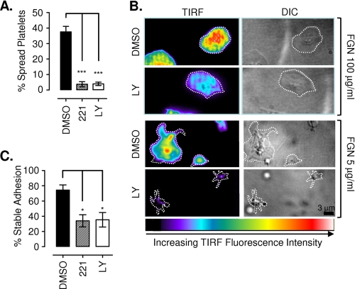
FIGURE 6.
PI3K plays an important role in strengthening high affinity integrin αIIbβ3 adhesive contacts. Washed human platelets were preincubated with vehicle alone (DMSO), LY294002 (LY; 25 μm), wortmannin (100 nm), or TGX221 (221; 0.5 μm) prior to stimulation with 1 unit/ml thrombin and application to immobilized fibrinogen (0.2–100 μg/ml). A, platelet-fibrinogen interactions under static conditions were recorded in real time for off-line analysis, as described under “Experimental Procedures.” Results are expressed as percentage of spread platelets and represent the mean ± S.E. of three independent experiments, with five random fields of view analyzed per experiment (***, p < 0.001). B, thrombin-stimulated DiIC12-labeled platelets were allowed to interact with an immobilized fibrinogen matrix (5–100 μg/ml) and observed using TIRF microscopy and DIC microscopy in real time as described under “Experimental Procedures.” Images were taken 10 min post-stimulation and are representative of three independent experiments. Note that the pseudocolor scale is representative of raw TIRF fluorescence intensity, with black/blue indicative of platelet membrane regions at the periphery of the evanescent field (∼100 nm from the coverslip surface) and red/white indicative of platelet membrane within ∼12–15 nm of the coverslip surface. C, thrombin-stimulated platelets were gently perfused (150 s−1) into fibrinogen-coated (2.0 μg/ml) microslides and allowed to interact for 5 min under static conditions prior to perfusion of Tyrode's buffer at 1800 s−1 for 5 min. The number of adherent platelets in the field of view before and after shear application was quantified, as described under “Experimental Procedures.” Results are expressed as percentage of stable adhesion (mean ± S.E., n = 3; *, p < 0.05).

