Figure 2.
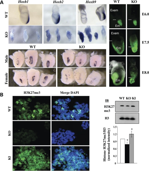
Huntingtin stimulated PRC2 during development. (A) Whole mount in situ hybridization Hoxb1, Hoxb2, Hoxb9 mRNA expression in E7.5 Hdhex/4/5/Hdhex/4/5 (KO) huntingtin null embryos was not properly silenced and restricted to the posterior, as observed for wild-type Hdh+/Hdh+ (WT) embryos (top). Decreased placental lactogen (PL-1) mRNA signal (purple) in bisected decidua revealed fewer giant trophoblast cells in female embryos lacking huntingtin (KO), compared with null male (KO) or wild-type (WT) embryos of either gender (bottom). Female embryos, at the indicated developmental stages (ectoplacental cone, Reichert's membrane removed), show aberrant Xp GFP-transgene reactivation in the Hdhex/4/5/Hdhex/4/5 (KO) extraembryonic tissue (Exem), compared with Hdh+/Hdh+ (WT) tissue, while random inactivation in the embryo proper (Em) appeared normal (right). (B) Confocal images of immunostained sections of day 4 Hdh+/Hdh+ (WT) and Hdhex/4/5/Hdhex/4/5 (KO) huntingtin null EBs revealed decreased histone H3K27me3 signal in the latter, whereas knock-in Hdh+/HdhQ111 (KI) EBs, expressing huntingtin with a 111 residue polyglutamine segment exhibit increased histone H3K27me3 signal. Immunoblot of nuclear extracts and plot of histone H3K27me3/H3 band intensity ratio, normalized to wild-type (white bar), confirmed significantly (P < 0.016) decreased histone H3K27me3 in Hdhex/4/5/Hdhex/4/5 (KO) extracts (black bar) and significantly (P < 0.044) increased histone H3K27me3 in knock-in Hdh+/HdhQ111 (KI) extract (grey bar), compared with wild-type (n = 4).

