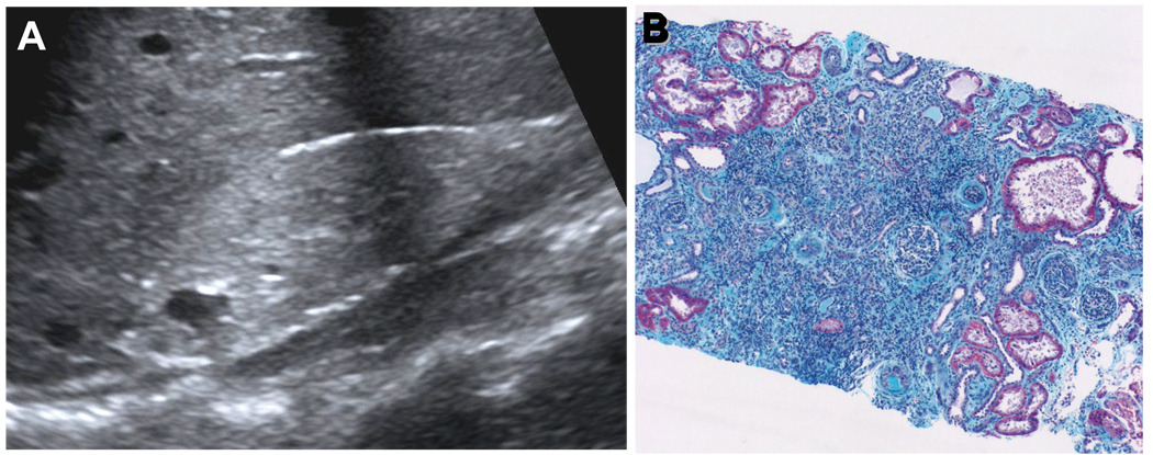
Figure 1. Morphology of nephronophthisis.
(A) Renal ultrasound demonstrates increased echogenicity, loss of corticomedullary differentiation, and the presence of corticomedullary cysts. In contrast to polycystic kidney disease kidneys are not enlarged. (B) Renal histology in NPHP shows the characteristic triad of renal tubular cysts, tubular membrane disruption, and tubulointerstitial cell infiltrates with interstitial fibrosis and periglomerular fibrosis (B is courtesy of D. Bockenhauer, London).

