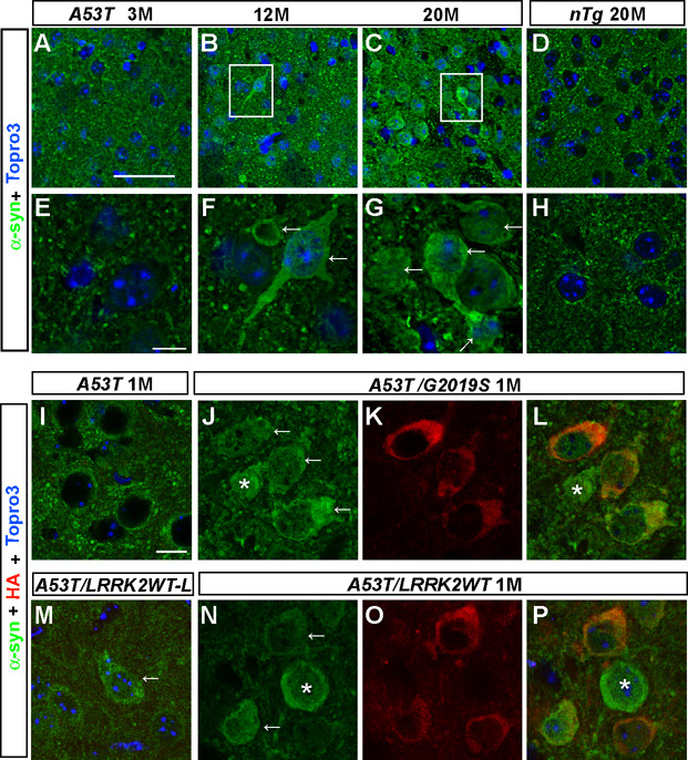
Figure 4. LRRK2 accelerates somatic accumulation of A53T α-syn in neurons.
(A–H) Representative images show α-syn staining (green) in striatal neurons of A53T mice at 3 (A), 12 (B), and 20 months of age (C), and nTg mice at 20 months of age (D). F and G represent enlarged images with the white boxes in B and C. Nuclei were stained with Topro 3 (blue). Scale bars: 50 µm (A–D); 10 µm (E–H).
(I–P) Representative images reveal α-syn staining (green) in striatal neurons of 1-month old A53T (I), A53T/G2019S (J, L), A53T/LRRK2WT-L (M), and A53T/LRRK2WT (N, P) mice. Human LRRK2 was stained with an anti-HA antibody (red, K–L, O–P). Nuclei were stained with Topro 3 (blue). Scale bar: 10 µm.

