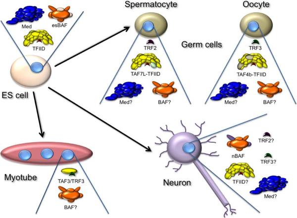
Figure 2. Model of Cell-Type-Specific Changes in General Transcription Factors.
ES cells and proliferative progenitors are believed to contain canonical TFIID consisting of TBP (red) and 12–15 associated TAFs (yellow), Mediator (blue), and ES BAF consisting of both common (orange) and ES cell-specific subunits (tan). Myotubes may replace TFIID with a novel complex of TRF (green) and TAF3 (yellow), and also downregulate Mediator and presumably retain a general BAF complex (orange). Neurons are known to contain a novel BAF complex with conserved (orange) and neuron specific subunits (purple). The disposition of Mediator and TFIID in most neuronal cell types is not known. Spermatocytes have elevated levels of TRF2 (purple) and of the TAF7 paralog TAF7l (lime) which may or may not be a component of an altered TFIID. Ovarian cells have elevated levels of TRF3 (green) and of an altered TFIID containing one or more subunits of the TAF4 paralog, TAF4b (tan). The subunit composition of Mediator and BAF complexes in germ cells is currently unknown.

