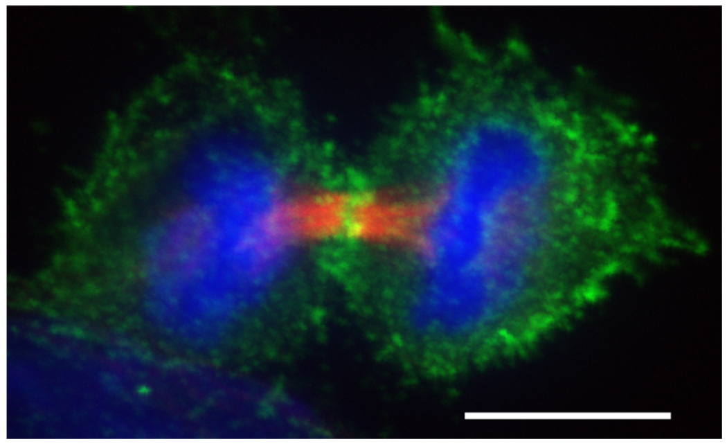
Figure 3. Dividing HeLa cell during early cytokinesis.
Phosphorylated myosin II accumulates at the cleavage furrow.  (red) were stained with anti-tubulin (Sigma T6199),
  (green) was stained with Phospho-Myosin Light Chain 2 (Ser19) antibody (Cell Signalling, 3671L),
  (blue) with DAPI. Scalebar represents 10 µM.

