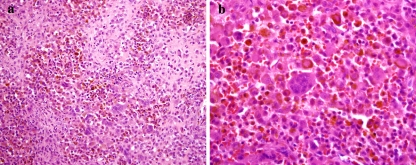
Fig. 2.
(a) Chondroblastoma of the temporal bone. The tumor contains pink nodules of chondroid differentiation surrounded by multinucleated giant cells and epithelioid tumor cells with brown pigment. (b) Higher magnification showing epithelioid cells with abundant eosinophilic cytoplasm containing brown hemosiderin

