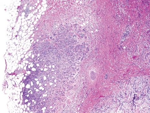
Fig. 1.
Minimally invasive carcinoma ex pleomorphic adenoma. The pleomorphic adenoma component with sclerosis is seen on the right, and the minor low grade carcinoma component infiltrates the surrounding adipose tissue. This carcinoma was immunophenotypically a myoepithelial carcinoma (stains not shown)

