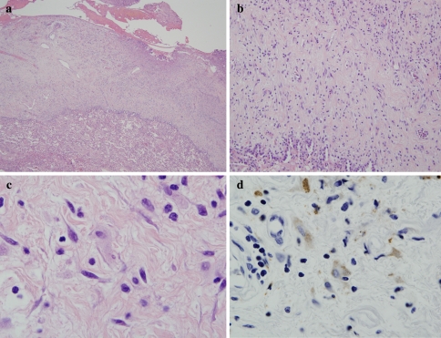
Fig. 3.
In Erdheim Chester disease, the basic process is one of a non-Langerhans cell histiocytopathy associated with fibrosis. The biopsy is of lung showing marked thickening of the pleura largely attributable to collagen deposition (a and b). Higher power magnification reveals scattered histiocytes (c) which can be highlighted with a CD68 stain (d)

