
Fig. 4.
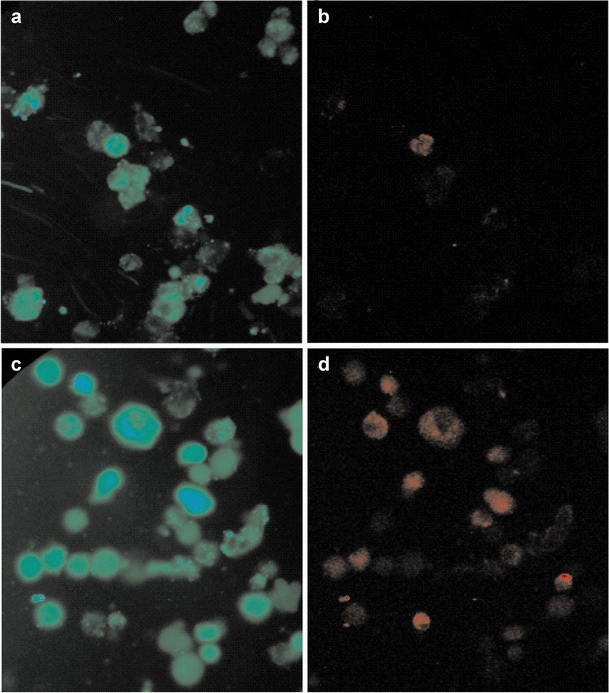
Images of immunofluorescence labelling of control cells (a, b) and cells treated with 200 mM AraC (c, d) at 24 h. When the green fluorescing label CFDA (a, c) is used, viable cells can be identified, whereas the red fluorescing label AnnV (b, d) is specific for apoptotic cells. At 24 h, 5% of the control cells have become apoptotic, while, after treatment with 200 mM AraC, 61% of cells are apoptotic