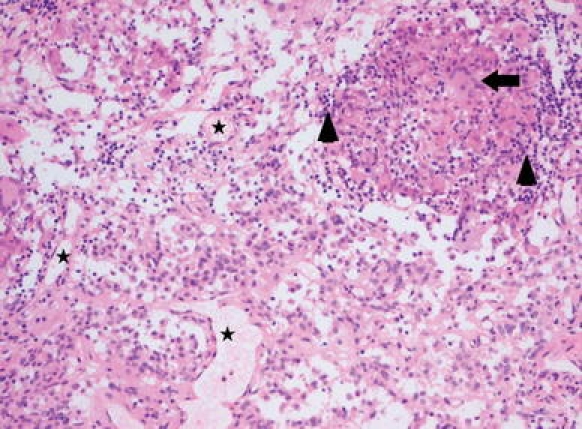
Fig. 1.

Patient No. 2. Pituitary null cell adenoma combined with granulomatous inflammation due to neurosarcoidosis. The pituitary adenoma consists of highly vascularized tissue (asterisks in some vessel lumina) and a monomorphous population of epitheloid tumor cells with eosinophilic cytoplasm; in the upper right hand corner a non-necrotizing granuloma is present consisting of histiocytes, some multinucleate giant cells (example indicated by arrow) and collections of small lymphocytes (arrowheads). Hematoxylin & Eosin staining ×100
