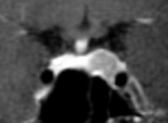
Fig. 7.

Patient No. 8. Preoperative coronal T1-weighted MRI demonstrating a sellar lesion attached to the left cavernous sinus, stretching the pituitary gland to the right and slightly deviating the pituitary stalk. Given the clinical presentation, the lesion was considered to be a GH-secreting pituitary adenoma
