
Fig. 4.
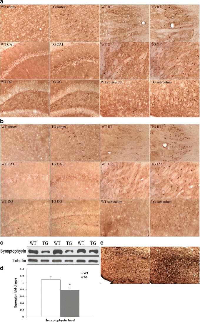
Molecular changes in COL25A1 TG mouse brains. a, b Immunohistochemical staining shows greater BACE1 (a) and p35/p25 (b) immunoreactivity in cortex, CA1, DG, reticular thalamic nuclei (RT), globus pallidus (GP), and subiculum of TG mice than WT littermates. c, d Western blot analysis of forebrain extracts shows synaptophysin loss in TG mice. Values are mean ± SEM; * p < 0.05 (t test). e Immunohistochemical staining of brain sections with anti-GFAP antibody shows astrocyte activation in the subiculum of TG mice