
Figure 2. Fluorescence images of mesenchymal cell growth.
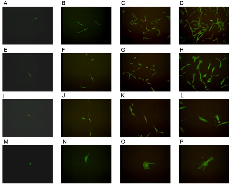
Images of EGFP expressing cells obtained on day 1 (A, E, I, M) post-sorting, day 3 (B, F, J, N), day 6 (C, G, K, O), and day 9 (D, H, L, P). Four distinct cell populations derived from single mesenchymal cells included high proliferative potential-mesenchymal colony forming cells (HPP-MCFC; A–D), low proliferative potential-mesenchymal colony forming cells (LPP-MCFC; E–H), mesenchymal cell clusters (MCC; I–L), and mature mesenchymal cells (MMC; M–P) which were observed over time in culture. Images shown in each row were taken from the same well over time.