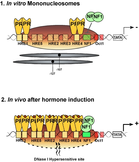
Figure 1. Schematic representation of the main cis elements in the MMTV promoter and their occupancy in nucleosomes assembled in vitro (upper panel) and in intact cells after hormone induction (lower panel).
The positions covered by the main population of histone octamers are indicated by the grey ovals. The HREs, the NF1 binding site and the TATA box are indicated. The numbers refer to the distance in nucleotides from the transcription start site. The hormone receptor (PR) dimers are depicted in yellow and the NF1 dimer by green circles.

