Figure 5.
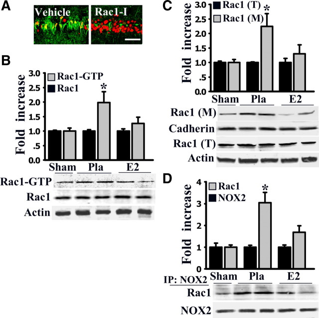
Critical role of Rac1 in ischemic neuronal damage in CA1 region and the effects of E2 in regulating Rac1 activation, membrane localization, and interaction with NOX2. A, Pretreatment with Rac1 inhibitor before ischemia resulted in significant neuroprotection as evidenced by an increased number of surviving neurons in the medial CA1 region. Red, NeuN; green, Fluoro-Jade B. B, Activated Rac (Rac1–GTP) was extracted from samples at 3 h reperfusion using glutathione S-transferase–p21-activated kinase and visualized by blotting with anti-Rac1 antibody. Total Rac1 and actin before extraction were determined as loading controls. C, Western blot analyses of membrane-bound Rac1 protein expression after 3 h reperfusion. Membrane localized Rac1 [Rac1 (M)] was significantly increased in the CA1 region by ischemia compared with sham rats, an effect that was significantly inhibited by E2 treatment. Total Rac1 protein [Rac1 (T)] was not changed by ischemia or E2 treatment. D, Protein samples from sham, Pla, and E2 groups at 3 h reperfusion were IP with anti-NOX2 antibody and blotted with anti-Rac1 and NOX2 antibodies. Complex formation between NOX2 and Rac1 was significantly increased in Pla group after ischemia and attenuated by E2. In B–D, values are means ± SE of five or six rats in each group. *p < 0.05 versus sham or E2 group.

