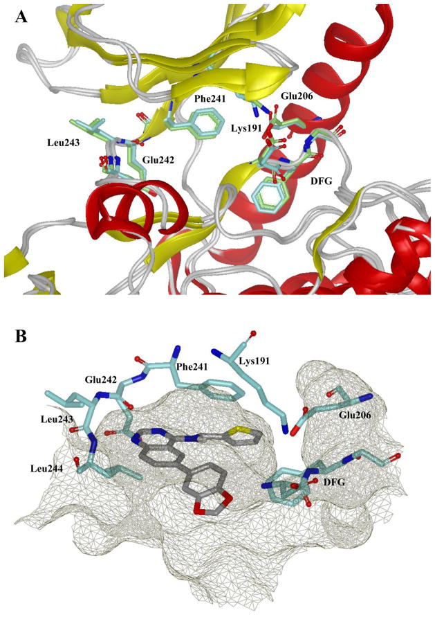
Figure 5.
A. Ribbon representation of the catalytic clefts in the Clk1 and Clk4 kinase domain. Clk4 kinase domain is a homology model derived from the X-ray structure of Clk1 (PDB code: 1Z57). Protein kinase structural elements are labeled and the key residues are colored: Clk1 in cyan, Clk4 in green. This figure was prepared with the program VIDA (OpenEye Scientific Software). B. Docking model of 4 in the Clk1 catalytic cleft. The binding pocket is depicted by molecular surface in mesh grey and the hydrogen bonds are labeled as green dotted lines. This figure was prepared with the program VIDA (OpenEye Scientific Software).

