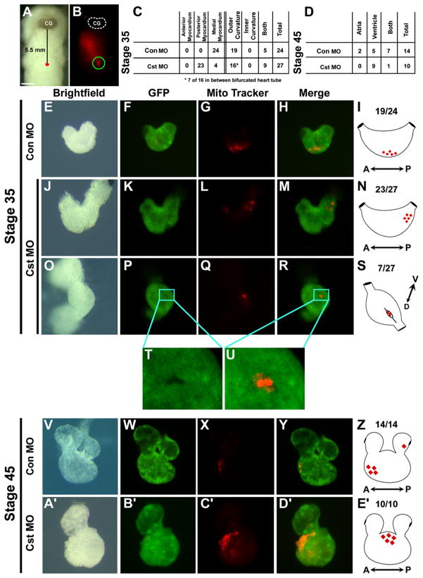
Figure 4. Fate Mapping Cardiac Ventral Midline Cells.
(A) Bright-field image of a living cardiac actin-GFP transgenic embryo injected with MitoTracker at Stage 29 along the ventral midline 5.5 mm posterior to the cement gland (CG). (B) Fluorescent image of the same embryo demonstrating the location of incorporated MitoTracker into cells at the ventral midline (ventral views with anterior to the top). Fluorescence anterior to site of injection is reflection off the surface of the live embryo. (C and D) Tabulation of the location of MitoTracker-labeled cardiac cells of control MO and CST-depleted embryo at (C) midtailbud Stage 35 and (D) tadpole Stage 45. Images of CA-GFP transgenic control MO-injected and CST-depleted (E–H, J–M, and O–U) Stage 35 and (V–Y and A′–D′) Stage 45 dissected hearts. (F, K, P, T, U, and Z) Corresponding images of GFP expression. (G, L, Q, X, and C′) Corresponding images of fated MitoTracker-labeled cardiac ventral midline cells. (H, M, R, U, Y, and D′) Merged images of GFP and fated MitoTracker-labeled ventral midline cells. (T and U) Note the fated ventral midline cells in a pocket of undifferentiated (GFP-negative) cardiomyocytes. (I, N, S, Z, and E′) Schematics representing fate of the cardiac ventral midline cells to the outer curvature of the ventricle in (I) Stage 35 and (Z) Stage 45 control MO-injected hearts. CST-depleted fated ventral midline cells located in the (N) posterior midline or (S) in an undifferentiated cleft in the outer ventricular myocardium in Stage 35 CST-depleted hearts and (E′) in a condensed mass of cells on the outer ventricle in Stage 45 CST-depleted hearts.

