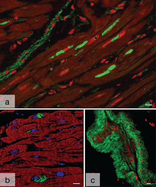
Figure 4).
Nonsarcomeric alpha (α)-actinin in human myocardium. A Several deposits of smooth muscle α-actinin (ACTN-1) (green) are in close proximity to the nucleus (red) in a longitudinal section of failing myocardium with dilated cardiomyopathy (red). Myocytes are dark. Note the labelling of blood vessels (top left). B ACTN-1-positive granules (green) are in close proximity to the nuclei (blue). Myocytes are red with phalloidin. Shadow projection in the confocal microscope. C Nonsarcomeric α-actinin in a tangentially sectioned arterial blood vessel. Faint staining of endothelial cells and strong labelling of the smooth muscle cells of the media (green). Nuclei are red. Bars represent 10 μm

