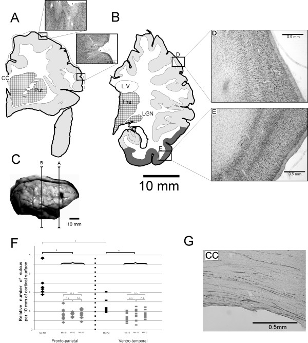
Figure 2.
Reconstructions of frontal Nissl-stained sections in PMG monkey. Panels A and B: Location of cortical malformation on coronal sections in the right hemisphere at two rostrocaudal levels. Sub-cortical structures, such as the thalamus (Thal.), the lateral geniculate nucleus (LGN), the putamen (Put) or the corpus callosum (CC) are visible and they do not present conspicuous abnormalities, but the lateral ventricle (L.V.) is enlarged, including two enlarged photo micrograph of the cortex showing the microscopic cortical ectopic sulci (#), where the layer I is clearly distinguishable. Light grey: unlayered cortex. Dark grey: normally layered cortex: Squared: subcortical nuclei Panel C: Photograph of the cerebral cortex in Mk-PM with the position of the sections depicted in panels A and B. Panels D and E: The cortical organization in layers is lost in the parietal lobe (D) but normal in the inferior temporal lobe (E). Panel F: Diagram showing the distribution of the number of sulci per cortical length (number of sulcus per 10 mm) in the PMG monkey (black) and in the three control monkeys, Mk-IU, Mk-I2 and Mk-IZ (grey). Diamonds represent the results obtained in the frontoparietal region and bars in the ventro-temporal region. N.s is for non-statistically significant. Panel G: Photomicrograph of the Corpus Callosum (CC) in the right hemisphere of Mk-PM showing large quantity of BDA stained fibers. Scale bar: 500 microns.

