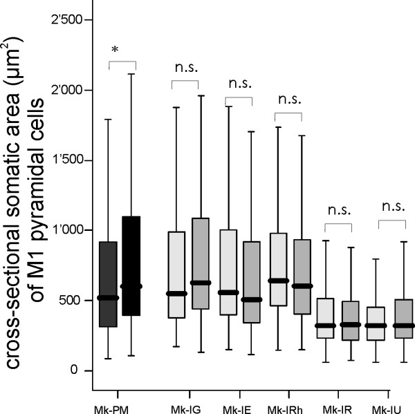
Figure 5.
Somatic cross-sectional areas of SMI-32 positive neurons in layer V in motor cortex. Box and whisker plots showing the distribution of somatic cross-sectional areas of SMI-32 positive neurons in layer V in motor cortex for the PMG monkey (dark grey) and four normal monkeys (light grey). In the box and whisker plots, the thick horizontal line in the box corresponds to the median value, whereas the top and bottom of the box are for the 75 and 25 percentile values respectively. The top and bottom extremities of the vertical lines on each side of the box are for the 90 and 10 percentile values, respectively. Mk-PM exhibited a significant inter-hemispheric difference of cross-sectional soma area (*p < 0.0001; Mann and Whitney test) whereas, in the normal monkeys, the difference was not statistically significant (n.s. p > 0.05; Mann and Whitney test). For each monkey, the left box and whisker plot corresponds to data from the left hemisphere.

