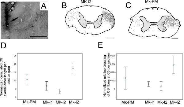
Figure 6.
Corticospinal projections in PMG monkey. Panel A: Site of BDA injection in M1 hand area of the left hemisphere (arrow), close to identified layer V pyramidal neurons. Panels B and C: Reconstructions of BDA stained corticospinal (CS) fibers in coronal sections of the cervical spinal cord at the C5 level, as a result of BDA injection in the right motor cortex of a normal monkey (B) and in the left motor cortex of the PMG monkey (C); scale bar 1 mm. For better visual comparison of both reconstructions, the reconstruction in panel C was drawn with the left spinal side on the right side of the drawing. Grey dots indicate the location and distribution of the CS axons in the white matter. In both monkeys, most fibers were found in the dorsolateral funiculus (DLF) contralateral to the injection site, the rest running along the dorsolateral and ventral funiculi ipsilateral to the injection site. In comparison to normal monkeys, slightly fewer CS fibers were found in the grey matter ipsilateral to the injection side in the PMG monkey. Panel D: Normalized cumulated axonal arbor length of corticospinal projections in the cervical grey matter in the PMG monkey (black) and in three normal monkeys (grey). Panel E: Number of midline crossing CS fibers at cervical level C5. The number of fibers crossing the midline was normalized by dividing it by the total number of labelled CS fibers present in the white matter (see methods for detail).

