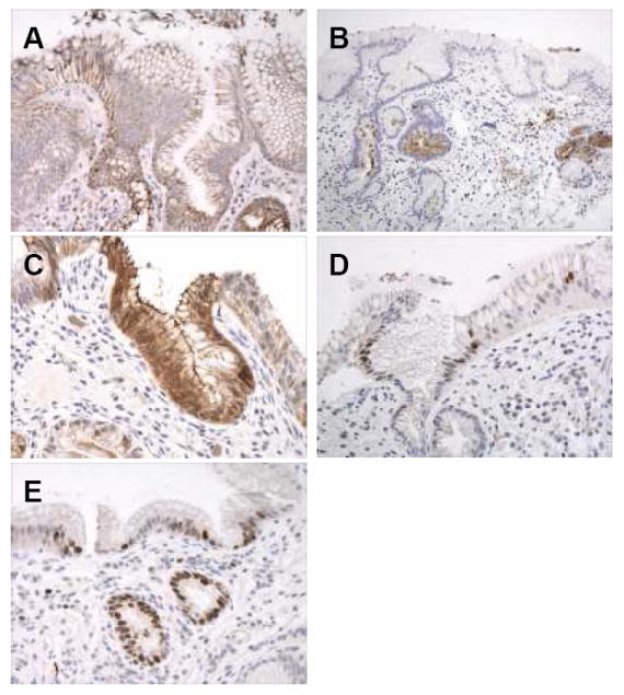
Figure 2.

(A-E): Immunohistochemical results of intestinal markers and Ki67 in a patient with columnar metaplasia of the esophagus, but without goblet cells. A. MUC-5AC showing diffuse cytoplasmic reactivity in surface and crypt epithelium. B. DAS-1 reactivity in columnar mucous cells within the deep portions of the crypt epithelium and within mucosal glands. C. Strong reactivity for villin in mucinous columnar cells in the surface and crypt epithelium. D. Nuclear staining for CDX2 in scattered cells in the surface and crypt epithelium. E. Nuclear Ki67 staining in the surface and crypt epithelium in an area of mucosa without active inflammation or ulceration.
