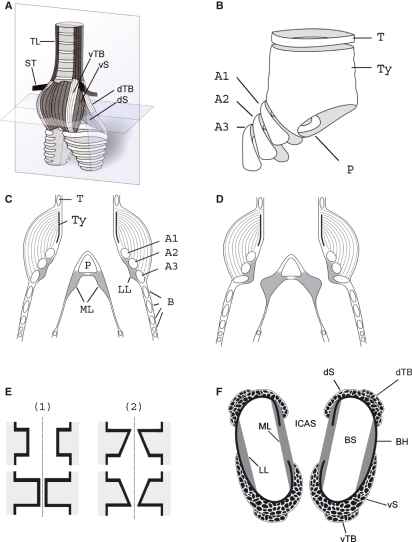
Fig. 1.
The songbird syrinx. (A) Schematic external ventral view of the excised organ. TL, tracheolateralis muscle; ST, sternotrachealis muscle; dTB and vTB, dorsal and ventral tracheobronchial muscle; dS and vS, dorsal and ventral syringeal muscle. (B) Schematic view of the cartilage framework of the syrinx (modified after Ames, 1971). The drum [or tympanum (Ty)] is located at the caudal end of the trachea. Its shape varies with species. A median dorso-ventral bar [pessulus (P)], spanning the lumen of the trachea, marks the end of the trachea and the beginning of the primary bronchi. The pessulus is one important attachment for the ML. A1–A3, bronchial half rings; T, tracheal ring. (C) Schematic of a frontal section of the syrinx (level indicated by the vertical plate in A). B, bronchial rings. (D) Schematic of the adduction setting of the ML and LL. (E) Simulating the labia oscillation assumes two major movement components: (1) a latero-lateral component and (2) a cranio-caudal component. The latter leads to a repeated change between a convergent and divergent cross-sectional profile. This out-of-phase movement of the upper and lower part of the labia produces larger asymmetries in the forces acting on the labia in the opening and closing phase of the oscillation than the inward and outward movement alone, resulting in a positive pressure on the labia during the opening phase and a net energy input to the labia maintaining a self-sustained oscillation. (F) Schematic horizontal section through the syrinx (level indicated by the horizontal plate in A). BS, bronchial lumen; BH, bronchial half ring.

