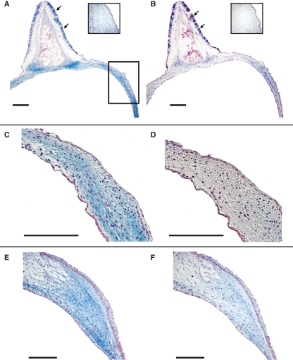
Fig. 3.
ML. (A,C,E) Alcian blue stain. (B,D,F) Hyaluronidase digestion and Alcian blue stain, same specimens as in A,C,E, respectively. (A–D) WCS. (E and F) ZF. The insets in A and B are control stains for Alcian blue stain and hyaluronidase digestion and Alcian blue stain. The square in A indicates the location of the higher magnification images in C–F. Goblet cells, located in the ciliated epithelium of the semilunar membrane, are indicated by arrows. Bars: 100 μm.

