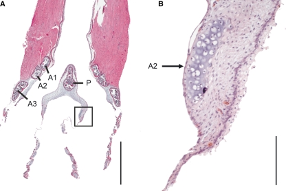
Fig. 7.
Frontal section from a ventral aspect of a ZF syrinx. The bronchial half ring arches into the ML ventrally and dorsally (see also Fig. 1F). The square in A indicates the area of magnification in B. A1, A2 and A3, first, second and third bronchial half ring, P, pessulus. Bars: in A: 1 mm; in B: 100 μm.

