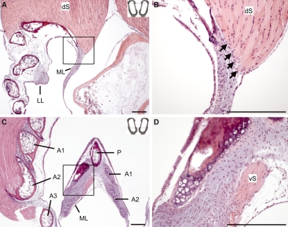
Fig. 8.
Two sections from a WCS syrinx indicating direct muscle attachments on labial tissue. The insets at the top right corners of A and C indicate the sectioning level along the dotted line (compare Fig. 1F). (A) Section through the dorsal part of the syrinx. The square in A indicates the area of magnification in B. Note that some fibers of the dorsal syringeal muscle (dS) attach directly to the ML (indicated by arrows). (C) Section through the ventral part of the syrinx. (D) Magnification of the medial aspect of the ML (as indicated by a square in C). The ventral syringeal muscle (vS) attaches to the ML. A1, A2 and A3, first, second and third bronchial half ring; P, pessulus. Bars: 100 μm.

