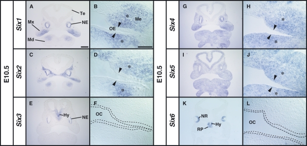
Fig. 1.
Localization of Six1, Six2, Six4 and Six5 mRNAs in the craniofacial region of the E10.5 mouse embryo before thickening of the dental epithelium. (B,D,F,H,J,L) High magnification images of the oral region of serial sections shown in A, C, E, G, I and K, respectively. (A,B) Expression of Six1 in the mesenchyme (Me) in the proximal region of maxillary (Mx) and mandibular (Md) arches (asterisk). No expression of Six1 in the oral epithelium (arrowhead). (C,D) Expression of Six2 in the Me of the maxillary and mandibular arches. (E,F) The expression of Six3 in the hypothalamus (Hy) and nasal epithelium (NE). The area within the broken line in F corresponds to the oral epithelium. (G,H) Expression of Six4 in the proximal mesenchymal cells (asterisk) and the oral epithelium (arrowhead) in the Mx and Md. (I,J) Uniform expression of Six5 in the craniofacial regions including the oral epithelium (arrow) and the mesenchyme (asterisk). (K,L) Expression of Six6 in the neural retina (NR), Hy and Rathke’s pouche (RP). Te, telencephalon; OE, oral epithelium; OC, oral cavity. Scale bars = 400 μm (A,C,E,G,I,K), 200 μm (B,D,F,H,J,L).

