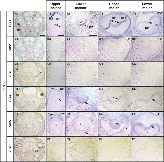
Fig. 4.
Localization of Six1, Six2, Six4 and Six5 mRNA on frontal sections at the bell stage (E18.5). (A1,B1,C1,D1,E1,F1) Low magnification images of expression patterns of the Six family genes at the anterior level including first molar tooth germs. (A1–A5) Expression of Six1 in the inner enamel epithelium (IEE) of the upper incisor tooth germ (arrow in A2,A3) and the IEE in the molar tooth germs (arrow in A4,A5). The expression of Six1 is detected in the dental follicle (DF) of the incisor and the molar tooth germs (arrowhead in A2,A4,A5). (B2–B5) Expression of Six2 in the DF (arrowhead in B4,B5) in the molar but not in the incisor tooth germs (B2,B3). (C2–C5) No expression of Six3 in the tooth germs. (D2–D5) Expression of Six4 in the IEE of the upper incisor and the molar tooth germs (arrow in D2–D5). (E2–E5) Expression of Six5 in the IEE of the incisor tooth germs (arrow in E2,E3) and the IEE and DF of the molar tooth germs (arrow and arrowhead in E4,E5). Asterisk indicates the expression of Six5 in the DF. (F2–F5) No expression of Six6 in the tooth germs. Md, mandible; Mx, maxilla; Tg, tongue; NC, nasal cavity; NR, neural retina. Scale bars = 400 μm (A1,B1,C1,D1,E1,F1), 200 μm (A2–A5,B2–B5,C2–C5,E2–E5,F2–F5).

