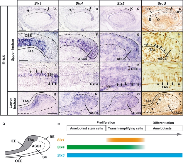
Fig. 5.
Comparative analysis of Six1/Six4/Six5 expression and the distribution of BrdU-positive cells in the incisor tooth germs along the anterior–posterior axis at E18.5 using serial sagittal sections. (A–D) In the upper incisor tooth germ, the expression of Six1 and Six4 is restricted to the posterior inner enamel epithelium including BrdU-incorporated cells (D). Six1 and Six4 signals are not detected in the BrdU-negative anterior inner enamel epithelium (arrowhead in A,B). Six5 is detected in the enamel epithelium and mesenchymal cells. (E–H) High magnification images of the cervical loop of the upper incisor tooth germ shown as in A–D. Six1 expression is detected in the posterior enamel epithelium (E) corresponding to the area in which BrdU-labeled transit-amplifying cells (TAs) reside (arrowheads in H). Six4 is expressed in the posterior region including both TAs and ameloblast stem cells (ASCs) (F,H). (I–L) Six1 and Six4 were expressed in the epithelium (arrowheads in I,J) including BrdU-labeled TAs (arrowheads in L) and become decreased in the anterior region. Six1 expression is also detected in the stratum intermedium (SI) (arrows in I). Six5 is detected in the outer enamel epithelium (OEE), inner enamel epithelium (IEE) (arrowheads in K), and the odontoblast (K). (M–P) In the cervical loop of the lower incisor tooth germ, Six1 expression is restricted in TAs (M,P). Six4 and Six5 are detected in TAs and the stellate reticulum in which ASCs reside (N,O,P). (Q) Localization of TAs and ASCs in the labial cervical loop of the lower incisor. (R) Time course of Six1/Six4/Six5 in the incisor tooth germs. E, enamel; O, odontoblasts; BE, basal epithelium. Scale bars = 100 μm (A–I), 200 μm (E–L), 100 μm (M–P).

