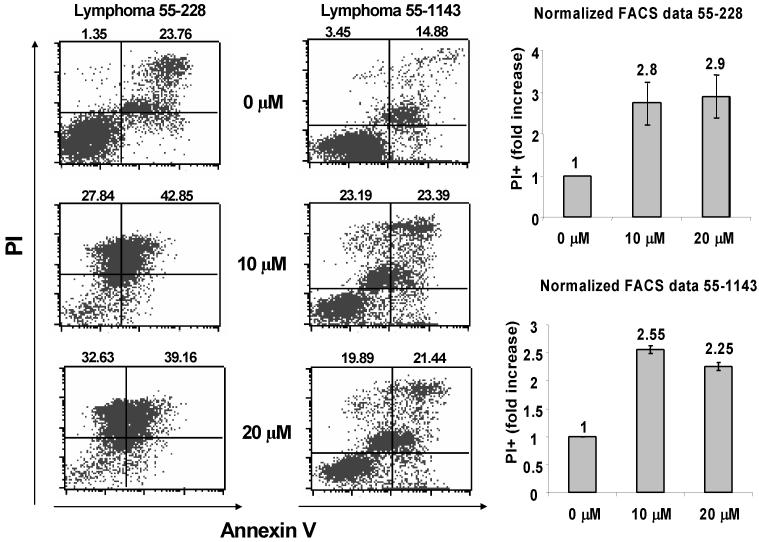
Fig. 3. Thymic lymphoma cells from Lck-MyrAkt2 mice are sensitive to GSK690693-induced apoptosis.
Tumor cells from two different mice were treated for 24 hrs with 0, 10 or 20 μM GSK690693 and then double labeled with propidium iodide (PI) and annexin V for flow cytometry analysis. Representative panels of the FACS analysis of lymphoma cells from mice 55–228 and 55–1143 are shown to the left. Bar graphs to the right illustrate 2–3 fold increase in PI-positive cells (dead tumor cells; upper quadrants of scatter plots) in each of the primary cultures in response to GSK690693. Error bars depict total standard deviation between replicate experiments.

