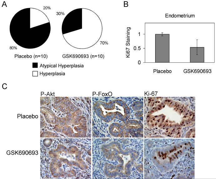
Fig. 4. GSK690693 delays endometrial tumor progression in Pten+/− knockout mice.
A, Pie charts depicting the percentage of mice that exhibited hyperplasia (20% in placebo vs. 70% in GSK690693-treated group) or that developed atypical hyperplasia (80% in placebo vs. 30% in GSK690693 group). Chi-Square test, p=0.03. B, Bar graph representing a significant difference between Ki-67 staining in placebo and GSK690693 mice (T-test, p=0.003) Stained and unstained cells from glandular endometrium from 5 fields of each of 5 placebo- and 5-GSK690693-treated mice were scored under a 40x objective (0.075 mm2 field), and percent positivity was normalized relative to staining in placebo mice. Error bars represent standard error. C, Immunohistochemical staining of endometrial neoplasms arising in Pten+/− knockout mice. Photographs were taken at 40x magnification. Like tumors from Lck-MyrAkt2 mice, P-Akt staining strongly localized to the cytoplasm and plasma membrane in placebo-treated mice, and was generally unchanged in treated mice. P-FoxO1/3 staining was cytoplasmic in placebo-treated mice (++ to +) and diminished (+ to +/−) in GSK690693-treated mice, and was sometimes nuclear.

