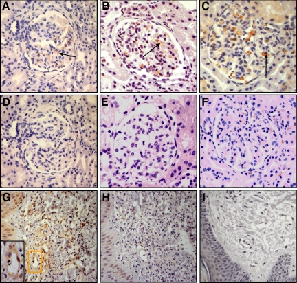
Figure 2.
IgA-binding regions of streptococcal M proteins in kidney samples from IgAN patients and kidney and skin samples from HSP patients. A: Renal cortex from a patient with IgAN labeled with anti-Sap60 (ie, antibodies to the IgA-BR of GAS serotype 60). Positive labeling is brown and noted in the mesangial area (see arrow). B: Renal cortex from a HSP patient labeled with anti-Sap4. C: Renal cortex from a HSP patient labeled with anti-Sap60. D: Renal cortex from the same patient as in A. Lack of labeling when the biopsy was incubated with preimmune rabbit serum. E: Normal renal cortex stained with anti-Sap60. F: Renal cortex from the same patient as in C. Specificity demonstrated by lack of labeling when the antibody was preincubated with its specific antigen (Sap60). G–H: Skin sample from a patient with HSP showing typical leukocytoclastic vasculitis with perivascular polymorphonuclear leukocyte and mononuclear cell infiltrates. G: Labeling with anti-Sap22 with pericapillary staining, magnified in the inset. H: Lack of reactivity when the primary antibody was preincubated with its specific antigen, Sap22, demonstrating specificity. I: Skin sample from a control strained with anti-Sap22. All panels counterstained with hematoxylin and shown at magnification × 400.

