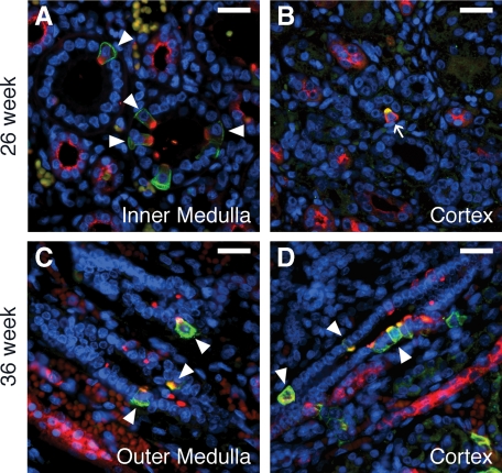
Figure 4.
ICs in the mid to late gestation human fetal kidney. In the 26-week kidney (A and B), type A ICs (arrowheads) predominate. Type B ICs (arrows) are first observed in the cortical CD at this age (B). In the 36-week gestation fetal kidney (C and D), a mix of type A (arrowheads), type B, and immature ICs is observed in both the outer medullary (C) and cortical (D) CD. Stains: red, vATPase; green, RhCG (A, C, and D) or pendrin (B); blue, DAPI. Scale Bars: A–D, 25 μm.

