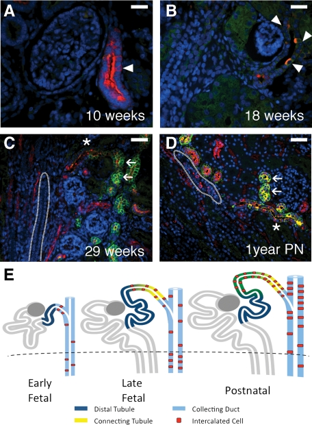
Figure 6.
The connecting tubule. At 10 weeks gestation, rare nephron segments can be observed resembling the distal tubule, which expresses uniform apical vATPase expression, but which contained isolated cells with stronger vATPase expression characteristic of ICs (arrowheads in A). These ducts may represent the earliest formations of the distal nephron and specifically, of the connecting tubule. By 18 weeks, ICs (arrowheads in B) became more distinct. By 29 weeks (C), and through 1 year postnatal development (D), the connecting tubule (asterisk) can be clearly identified as a transition segment between the distal tubule (arrows) and the CD (outline). The connecting tubule appears to represent a progressive overlapping of the mesenchymally-derived distal tubule and ureteric bud-derived CD with ICs derived from the latter, as depicted in (E). Stains: red, vATPase; green, RhCG; blue, DAPI. Scale bars: A–B, 25 μm; C–D, 50 μm.

