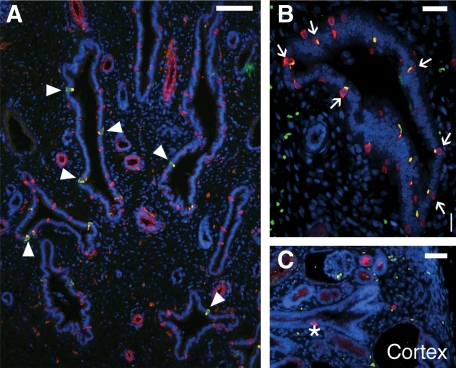
Figure 7.
ICs in the early obstructed human fetal kidney. At 18 weeks gestation, an increase in IC fraction can be observed (A and B). Furthermore, unlike the normal 18-week gestation kidneys, where immature ICs predominate, differentiated type A (arrowheads) and type B (arrows) ICs can be observed in medullary CDs of the obstructed kidney. ICs are also observed in the cortical CD (asterisk) near the nephrogenic zone highlighting premature differentiation of ICs in the region (C). Stains: red, vATPase (A and C) or pendrin (B); green, RhCG; blue, DAPI. Scale bar: A, 100 μm; B, 25 μm; C, 50 μm.

