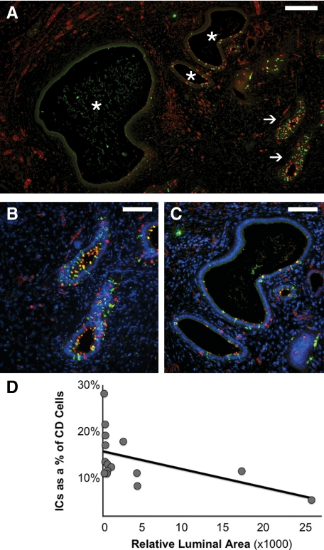
Figure 8.
ICs in the late gestation obstructed fetal kidney. As with the early gestation obstructed kidney, mildly affected CDs of the 36-week gestation obstructed fetal kidney (arrows in A, magnified in B) exhibit an increase in IC fraction. However, more severely dilated CDs displayed decreased IC abundance (asterisks in A, magnified in C). In the late gestation fetal kidney, injury and progressive dilation of the obstructed CD (measured as relative luminal surface area in pixels), correlated with a progressive decline in the relative number of intercalated cells in the CD (D; R2 = 0.255). Stains: red, vATPase; green, RhCG; blue, DAPI. Scale bars: A, 200 μm; B–C, 100 μm.

