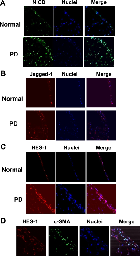
Figure 3.
Immunofluorescence evidence for the increased Notch signaling activation in fibrotic peritoneum. Peritoneal sections of normal rats or rats on 28-day PDF treatment (100 ml/kg) were paraffin-fixed and stained with antibodies against NICD (A), Jagged-1 (B), and HES-1 (C). D: Peritoneal sections of rats on 28-day PDF treatment (100 ml/kg) were paraffin-fixed and costained with antibodies against HES-1(red) and α-SMA (green). Nuclei were stained with DAPI (blue). Images (magnification ×400) were taken by confocal microscopy.

