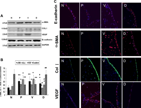
Figure 7.
DAPT treatment attenuated the expression of several protein characteristics of EMT and peritoneal fibrosis. A: The protein expression of α-SMA, collagen I, VEGF, and E-cadherin in peritoneum was examined by Western blot. B: Graphic representation of relative abundance of α-SMA, collagen I, VEGF, and E-cadherin normalized to GAPDH. Data are expressed as mean ± SD (n = 6). *P < 0.05 versus normal rats. **P < 0.01 versus normal rats. #P < 0.05 versus PDF-treated rats and vehicle treated rats. ##P < 0.01 versus PDF-treated rats and vehicle control rats. C: Immunoflurescence staining of α-SMA, collagen I, VEGF, and E-cadherin in peritoneum. Paraffin-fixed peritoneum sections were stained with indicated antibodies. Nuclei were stained with DAPI (blue). Images (magnification ×400) were taken by confocal microscopy. N indicates normal rats; P, PDF-treated rats; V, vehicle control rats treated with DMSO together with PDF. D, Rats treated with DAPT together with PDF.

