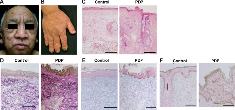
Figure 1.
Clinical findings and histology of a PDP patient. A, B: Clinical appearances of the face (A) and hands (B) in PDP (case 1) are shown. C: Histology of the face in PDP shows thickened dermis, and hyperplasia of the sebaceous and sweat glands by H&E staining compared with a healthy donor (control). Scale bars = 300 μm. D: The sample of PDP stained with Elastica van Gieson shows thick collagen and elastic fibers in the dermis compared with a healthy donor. E: The intensity of mucinous ground substance observed by Alcian blue staining is comparable between a healthy donor and a PDP patient. F: Melanocytes in the patient with PDP and a healthy control are identified with Fontana Masson staining. Scale bars = 100 μm (D−F).

